
Seeing Beyond the Cephalogram: What CBCT Reveals About the Airway
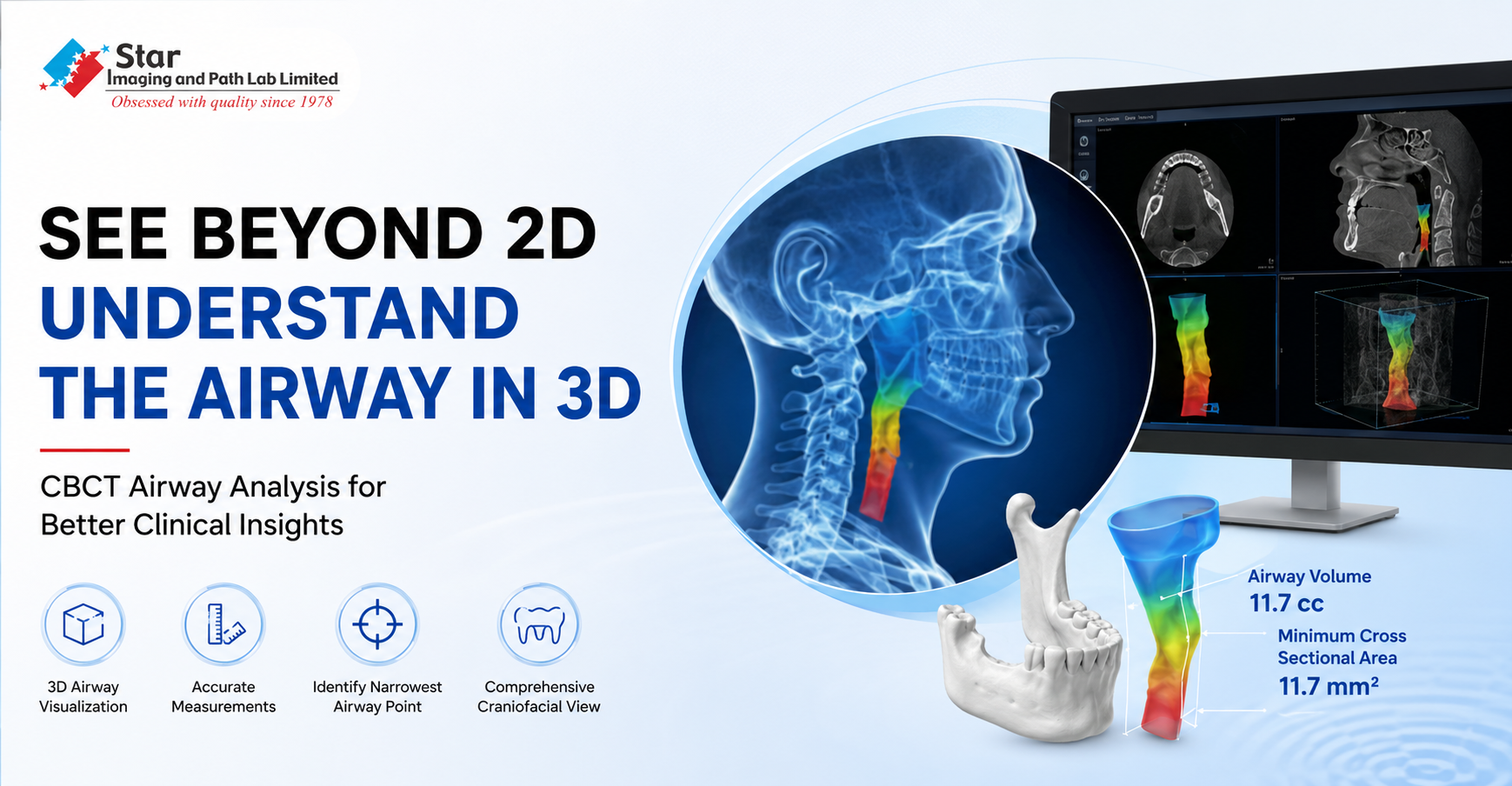
A cephalogram gives us a profile. The airway, however, has no profile. It has depth, width, changing contours and a narrowest point that may not tell its full anatomical picture from a single two dimensional projection.
That distinction becomes important when a patient presents with symptoms such as snoring or difficulty breathing during sleep. In such situations, CBCT, or Cone Beam Computed Tomography, can add another layer to craniofacial imaging by allowing the upper airway to be examined in three dimensions.
When the Airway Becomes Part of the Examination
A recent case reviewed by Dr. Sakshi Kamra, Consultant Maxillofacial Radiologist, involved a patient who complained of difficulty breathing while sleeping and snoring. The CBCT examination was undertaken as part of a broader craniofacial assessment, but the airway offered an important area for closer evaluation.
On three dimensional airway reconstruction, the airway could be separated from the surrounding anatomical structures and examined as a defined space. The colour coded rendering brought attention to a particular region where the airway became narrower.
The measurements added another perspective. The total airway volume was 11.7 cc, while the minimum cross sectional area measured 11.7 mm⊃2;. These figures describe the anatomy captured in this examination. They should not, however, be interpreted in isolation as diagnostic cut offs for obstructive sleep apnoea.

What a CBCT Airway Analysis Can Show
This is where CBCT airway assessment becomes particularly useful. Instead of relying on a single projected image, the clinician can examine airway volume and cross sectional dimensions at different anatomical levels. The analysis can also indicate the location of the narrowest segment, its anteroposterior and transverse dimensions, airway length and the shape of the airway lumen.
For a maxillofacial radiologist, these details are valuable because the airway does not exist independently of the surrounding anatomy. Its form can be assessed alongside the jaws, facial skeleton and adjacent structures, giving the clinician a more complete anatomical view.

Where CBCT Fits in Sleep Apnoea Assessment
There is an important distinction to make here. CBCT shows anatomy. Obstructive sleep apnoea is a physiological condition that occurs during sleep.
Airway dimensions can vary with factors such as head position, tongue position, respiration and other patient related variables. A CBCT examination captures anatomy at a particular point in time. It cannot reproduce the changes in airway behaviour that may occur during sleep.
This is why CBCT airway analysis should be viewed as an anatomical component of a wider clinical assessment rather than a standalone test for obstructive sleep apnoea. When OSA is suspected, appropriate sleep evaluation remains necessary for diagnosis and assessment of severity.
A Radiologist’s Perspective
After more than a decade in diagnostic imaging, Dr. Sakshi Kamra emphasises that the value of CBCT lies in interpreting the anatomy rather than reducing it to a measurement.

“A number such as airway volume or minimum cross sectional area becomes meaningful only when it is considered alongside the anatomy it represents. CBCT gives us the ability to locate and visualise areas of airway narrowing in three dimensions and to assess them in relation to the surrounding craniofacial structures. Its strength is anatomical detail, but that detail must always be interpreted within the clinical context.”
The Bigger Picture
The significance of this case is not that CBCT can answer every question about the airway. It cannot. Its value lies in answering a different and important question: what does the airway actually look like in three dimensions?
For selected orthodontic, dentofacial and maxillofacial evaluations, CBCT imaging can provide anatomical information that may be difficult to appreciate on conventional two dimensional radiography. When interpreted carefully, it gives clinicians another perspective on the relationship between craniofacial anatomy and the upper airway.
Frequently Asked Questions
Question: Can a CBCT scan diagnose sleep apnoea?
Answer: CBCT provides a three dimensional assessment of airway anatomy, but it cannot assess the dynamic airway changes that occur during sleep. Diagnosis of obstructive sleep apnoea is an adjunct with CBCT. But it requires appropriate clinical and objective sleep assessment such as sleep testing and polysomnography. Hence CBCT is an anatomical adjunct to OSA assessment.
Question: What does minimum cross sectional area mean in a CBCT airway analysis?
Answer: It represents the smallest measured cross sectional area along the assessed airway. In this case, the measurement was 11.7 mm⊃2;. It identifies the most constricted region within the analysed airway but should not be treated as an independent diagnostic threshold for OSA.
Question: Why can CBCT show information that a cephalogram may not?
Answer: A cephalogram presents anatomical structures as a two dimensional projection. CBCT permits assessment across multiple planes and reconstruction of the airway in three dimensions, allowing transverse dimensions, volume and the location of maximum narrowing to be evaluated.
Question: Can airway measurements on CBCT vary between patients or examinations?
Answer: Yes. Head posture, tongue position, respiratory phase, patient positioning and the method used for airway segmentation can influence measurements.
Question: When can CBCT airway assessment be useful?
Answer: It can be useful in identifying narrowing or obstructions in Airway, adenoid tonsillar enlargement, suspected OSA patients, craniofacial abnormalities, significant skeletal discrepancies. CBCT may also be valuable for treatment planning in Orthodontics and orthognathic procedures.


.jpg)
.jpg)
.jpg)
.png)
.gif)
.png)
.png)

Comments List