
Painful Scoliosis in Children Could Be More Than Just a Curved Spine
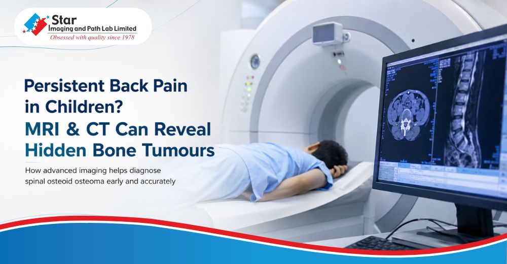
Back pain in children is often overlooked because it is commonly linked to poor posture, sports activities, or temporary muscle strain. However, when the pain becomes persistent, worsens at night, and improves after taking pain relieving medication, it deserves careful medical attention.
One such condition is osteoid osteoma, a small benign bone tumour that can produce severe pain despite its size. Although it is more commonly seen in the long bones of the arms and legs, it can also develop within the spine, where it often presents as painful scoliosis. Because the symptoms may resemble more common spinal conditions, advanced imaging plays an essential role in identifying the exact cause.
A recent case evaluated by Dr. Sameer Sood, HOD Radiodiagnosis, highlights why combining MRI and CT Scan provides the most accurate diagnosis in these situations.
When Persistent Night Pain Points Towards Osteoid Osteoma
A 13 year old boy reported persistent upper lumbar spine pain for nearly six months. The pain became more intense during the night and was significantly relieved after taking nonsteroidal anti inflammatory medication. There was no history of injury, fever, weight loss, or weakness in the legs.
Clinical examination showed tenderness over the second lumbar vertebra with mild muscle spasm around the lower back. While movement of the lumbar spine was slightly restricted because of pain, neurological examination remained completely normal.
The initial MRI of the lumbar spine demonstrated a focal abnormality involving the left pars interarticularis of the L2 vertebra. The lesion appeared dark on T1 weighted images and brighter on T2 weighted and STIR sequences. More importantly, MRI revealed extensive surrounding bone marrow oedema and inflammation within the adjacent soft tissues. There was no compression of the spinal canal or nerve roots.
Although MRI clearly demonstrated the inflammatory changes, the exact nature of the lesion became evident only after performing a thin section CT Scan of the lumbar spine.
CT Scan identified a well defined 8 millimetre nidus within the left pars interarticularis of L2. The lesion showed central mineralisation surrounded by dense reactive sclerosis, a classic appearance of osteoid osteoma. There was no evidence of vertebral instability, spondylolysis, or spondylolisthesis.
The combined findings from MRI and CT Scan confirmed the diagnosis of osteoid osteoma involving the posterior elements of the L2 vertebra.
Why MRI and CT Scan Complement Each Other in Spinal Osteoid Osteoma
Diagnosing osteoid osteoma of the spine can be challenging because symptoms often mimic mechanical back pain or other spinal disorders. In many patients, the lesion itself is extremely small, while the surrounding inflammatory reaction is extensive.
This is where advanced imaging becomes invaluable.
MRI is highly sensitive in demonstrating bone marrow oedema, soft tissue inflammation, and any effect on nearby neural structures. It provides a comprehensive assessment of the surrounding tissues but may occasionally make the small nidus difficult to appreciate because of the extensive inflammatory response.
CT Scan, on the other hand, remains the gold standard for identifying the characteristic nidus with central mineralisation and surrounding sclerosis. Thin section CT provides excellent anatomical detail, allowing precise localisation of the lesion and supporting treatment planning.
When both imaging techniques are interpreted together, they provide a complete understanding of the condition and significantly improve diagnostic confidence.
Persistent night back pain in children, particularly when relieved by anti inflammatory medication, should never be dismissed as simple muscular pain. While osteoid osteoma is a benign condition, timely diagnosis helps prevent prolonged discomfort, unnecessary investigations, and progressive spinal deformity such as painful scoliosis.
At Star Imaging and Path Lab Limited, advanced MRI and Multislice CT Scan technology, combined with the expertise of experienced radiologists, supports accurate diagnosis of complex spinal conditions, helping clinicians make well informed treatment decisions.

"Back pain in children should always be evaluated carefully when it is persistent, worse at night, or associated with spinal curvature. MRI and CT Scan together provide complementary information, enabling confident diagnosis of conditions such as osteoid osteoma that may otherwise remain undetected."
Dr. Sameer Sood
HOD Radiodiagnosis
Star Imaging and Path Lab Limited
Experiencing persistent back pain or have been advised an MRI or CT Scan of the spine?
At Star Imaging and Path Lab Limited, our advanced MRI and Multislice CT Scan services, supported by experienced radiologists, provide detailed evaluation of complex spinal conditions with a strong focus on diagnostic accuracy.
Book your appointment today and consult your referring doctor with confidence, backed by high quality imaging and expert interpretation.
Frequently Asked Questions
Question: Can osteoid osteoma cause scoliosis in children?
Answer: Yes. Osteoid osteoma involving the spine can produce painful muscle spasm that leads to painful scoliosis. Unlike structural scoliosis, the spinal curvature often improves once the tumour is treated.
Question: Why is back pain worse at night in osteoid osteoma?
Answer: Night pain is one of the hallmark features of osteoid osteoma because the tumour produces prostaglandins that stimulate pain receptors. The pain typically improves after taking NSAID medication.
Question: Is MRI or CT Scan better for diagnosing osteoid osteoma?
Answer: Both are important. MRI demonstrates inflammation and bone marrow oedema, while CT Scan provides the clearest visualisation of the nidus, making it the preferred imaging technique for confirming the diagnosis.
Question: Can osteoid osteoma be missed on MRI?
Answer: Yes. Extensive surrounding inflammation may sometimes obscure the small nidus on MRI. This is why CT Scan is frequently recommended when clinical suspicion remains high.
Question: When should persistent back pain in a child be investigated?
Answer: Back pain lasting several weeks, pain that becomes worse during the night, pain associated with scoliosis, or symptoms that repeatedly improve with NSAIDs should be evaluated with appropriate imaging under medical supervision.


.png)
_vs_Covid-19_How_the_Two_Viruses_Differ_and_Why_It_Matters.jpg)


.jpg)

.jpg)
.jpg)
Comments List