
When Ultrasound Reveals the Unexpected: A Rare Intra Abdominal Pregnancy That Changed Clinical Decision Making
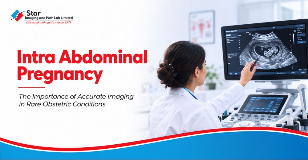
Every radiologist encounters cases that reaffirm one of the most important principles of medical imaging. Images do not simply document anatomy. They often answer questions that have not yet been asked.
One such case recently came under the care of Dr. Anisha Gupta, Consultant Radiologist, whose clinical experience spanning more than two decades has been built on careful observation, disciplined interpretation, and evidence based reporting.
What initially appeared to be a routine ultrasound referral for irregular menstrual cycles and abdominal pain evolved into the diagnosis of one of the rarest obstetric conditions encountered in modern radiology.

The patient presented with complaints that are not uncommon in everyday clinical practice. Irregular menstrual cycles accompanied by abdominal discomfort frequently prompt an ultrasound examination to evaluate the uterus, ovaries, and surrounding pelvic structures. However, this examination revealed an extraordinary finding.
The uterus was completely empty. Yet, a live fetus was clearly visualized outside the uterine cavity within the right adnexal region.
At that moment, the imaging findings immediately shifted the clinical perspective from a routine gynecological evaluation to a potentially life threatening obstetric emergency.
Understanding an Exceptionally Rare Pregnancy
An ectopic pregnancy occurs when a fertilized ovum implants outside the uterine cavity. More than ninety five percent of ectopic pregnancies occur within the fallopian tubes. Because the fallopian tubes cannot accommodate a growing pregnancy, these pregnancies generally become symptomatic during the early weeks of gestation and require prompt medical attention.
An Intra Abdominal Pregnancy represents one of the rarest forms of Ectopic Pregnancy.
In these exceptional situations, the pregnancy develops within the abdominal cavity instead of the uterus. Such cases account for only a very small proportion of ectopic pregnancies worldwide and present considerable diagnostic and surgical challenges because the fetus develops in close proximity to major abdominal and pelvic organs.
Unlike more familiar forms of ectopic pregnancy, an intra abdominal pregnancy may not always present with the expected clinical features. This makes careful Ultrasound Examination and advanced Diagnostic Imaging indispensable for arriving at the correct diagnosis.

The Critical Contribution of Ultrasound and MRI
The ultrasound examination served as the first indication that the clinical picture was far more complex than anticipated. Identifying an empty uterus despite the presence of a live fetus required careful anatomical evaluation and systematic assessment of the pelvis.
Recognising these unusual findings, an urgent MRI Scan was performed the very same evening.
The MRI confirmed the diagnosis of a Live Intra Abdominal Pregnancy, accurately demonstrating the location of the fetus while defining its relationship with adjacent pelvic and abdominal structures. This detailed anatomical information proved invaluable for the treating clinical team, allowing well informed surgical planning before intervention.
Rather than replacing ultrasound, MRI in Pregnancy complemented the initial findings by providing additional anatomical clarity that could not be completely appreciated on ultrasound alone.
Advanced Imaging That Supports Clinical Decision Making
At Star Imaging and Path Lab Limited, advanced diagnostic imaging is strengthened by experienced radiologists who understand that every examination has the potential to influence the next clinical decision.
Complex cases are evaluated through a combination of Advanced Ultrasound, High Resolution MRI, and comprehensive clinical correlation, enabling physicians to understand anatomy with greater precision whenever routine imaging reveals unexpected findings.

The purpose of imaging extends beyond producing high quality scans. It is about delivering clinically meaningful information that supports confident diagnosis, multidisciplinary collaboration, and appropriate treatment planning.
Whether evaluating neurological disorders, abdominal diseases, musculoskeletal conditions, oncological cases, or complex obstetric imaging, every examination is interpreted with the same systematic approach that prioritises diagnostic accuracy and clinical relevance.
Rare cases such as this reinforce why experienced radiological interpretation remains one of the most valuable components of modern healthcare.
Experience Shapes Interpretation
Modern imaging technology continues to advance, but technology reaches its full potential only when guided by clinical expertise.
Cases like this remind us that every scan deserves a disciplined, methodical review. Even referrals that appear routine can occasionally reveal diagnoses that challenge conventional expectations.
For a radiologist, the responsibility extends beyond identifying abnormalities. It involves recognising unexpected imaging patterns, communicating them clearly to the treating physician, and providing information that directly influences patient management.
"Technology captures anatomy. Experience recognises significance. In radiology, the difference between a routine examination and a life changing diagnosis often lies in the discipline to evaluate every image without assumption. Every scan deserves complete attention because uncommon diagnoses rarely announce themselves. They reveal themselves only to those willing to look carefully enough."
Dr. Anisha Gupta Consultant Radiologist

Why This Case Matters
Although an Intra Abdominal Pregnancy is exceptionally uncommon, it serves as an important reminder that imaging should always be interpreted through careful anatomical assessment rather than expectation alone.
The complementary use of Ultrasound and MRI transformed clinical suspicion into diagnostic certainty, allowing the treating team to proceed with clarity and confidence.
For radiologists, obstetricians, and clinicians alike, cases such as these reinforce the value of meticulous imaging, multidisciplinary collaboration, and evidence based clinical decision making.
Why Patients Trust Star Imaging and Path Lab Limited for Their Diagnostic Investigations
At Star Imaging and Path Lab Limited, we combine experienced radiologists with advanced diagnostic technology to deliver comprehensive imaging services across Ultrasound, MRI Scan, CT Scan, Digital X Ray, Mammography, PET CT, Doppler Studies, CBCT, FibroScan, and specialised women's imaging.
Every examination is performed with a commitment to clinical accuracy, detailed reporting, and meaningful diagnostic interpretation that supports both physicians and patients.
Whether your doctor has advised an MRI Scan, Ultrasound, or a second opinion on a complex imaging study, our specialists are equipped to provide comprehensive diagnostic support across Delhi NCR.
To book your appointment with Star Imaging and Path Lab Limited, contact your nearest Star Imaging centre today and experience advanced diagnostic imaging backed by clinical expertise.
Frequently Asked Questions
Question: What is an Intra Abdominal Pregnancy?
Answer: An Intra Abdominal Pregnancy is an exceptionally rare form of ectopic pregnancy in which the pregnancy develops within the abdominal cavity instead of inside the uterus. It requires specialised radiological assessment and multidisciplinary clinical management.
Question: How is an Intra Abdominal Pregnancy diagnosed?
Answer: Diagnosis usually begins with a detailed Ultrasound Examination. When imaging findings are unusual or complex, an MRI Scan provides additional anatomical detail, helping clinicians accurately determine the location of the pregnancy and its relationship with surrounding organs.
Question: Why is MRI performed after ultrasound in complex pregnancy cases?
Answer: While ultrasound often establishes the initial diagnosis, MRI offers superior soft tissue characterisation and a comprehensive anatomical overview. This information is invaluable when planning surgical or medical management in rare obstetric conditions.
Question: Can every ectopic pregnancy be identified during the early weeks?
Answer: Many ectopic pregnancies are diagnosed early, but exceptionally rare forms such as Intra Abdominal Pregnancy may occasionally progress further, making careful imaging interpretation essential whenever clinical findings do not correspond with routine expectations.
Question: What makes Intra Abdominal Pregnancy difficult to diagnose?
Answer: Its rarity, variable presentation, and unusual anatomical location can make diagnosis challenging. A systematic evaluation by an experienced radiologist, combined with advanced imaging modalities such as Ultrasound and MRI, is often critical for establishing the diagnosis.
Question: When should an MRI be considered after an ultrasound?
Answer: MRI is particularly valuable when ultrasound findings are inconclusive, when anatomical relationships require further clarification, or when detailed preoperative imaging is necessary to guide clinical decision making.


.png)


.png)
.jpg)

.png)

Comments List