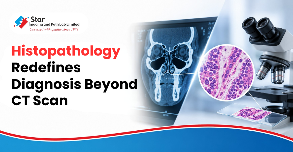
Histopathology Redefines Diagnosis Beyond CT Scan
Medicine often begins with observation. A patient presents with symptoms, imaging is performed, and the findings are interpreted within the framework of the most probable clinical possibilities. Most of the time, this approach is remarkably reliable. Occasionally, however, a tissue specimen arrives at the pathology laboratory and quietly changes the entire conversation.
This case serves as a powerful reminder that diagnosis is not built on probability alone. It is built on evidence.
The radiological evaluation suggested a complex but seemingly coherent picture. There was right lacrimal sac obstruction, extensive pansinusitis, an antrochoanal polyp, and findings that even raised the possibility of a fungal process. The lesion demonstrated soft tissue involvement with associated bony changes, making it a clinically significant abnormality requiring further evaluation.
Viewed in isolation, these findings appeared to support an inflammatory or infective pathology. The imaging told a convincing story.
Histopathology told a different one.
The Tissue Revealed What Imaging Could Not
Microscopic examination of the submitted specimen demonstrated malignant neoplastic cells arranged in sheets and nests. The cells exhibited marked pleomorphism with irregular nuclear membranes, prominent nucleoli, and appreciable mitotic activity. Areas of necrosis and accompanying inflammatory infiltrates were also identified within the tissue.
These are not merely technical observations confined to a pathology report. They are biological signatures that define how a lesion behaves. While radiology can identify a mass and describe its extent, histopathology determines its cellular identity and biological character.
Based on the microscopic findings, a provisional diagnosis of a poorly differentiated malignant neoplasm was made, with features favoring a poorly differentiated meibomian gland carcinoma. Immunohistochemistry (IHC) was recommended for definitive categorization and confirmation of the diagnosis.
The significance of this conclusion cannot be overstated. A diagnostic pathway that initially considered inflammatory and benign polypoidal lesions was fundamentally altered by cellular evidence obtained through tissue examination.
Why This Case Matters
Modern diagnostic medicine is often described as multidisciplinary, yet cases like this demonstrate why that collaboration is indispensable.
Computed tomography excels at defining anatomy. It identifies obstruction, evaluates the extent of disease, demonstrates bone involvement, and provides surgeons with an anatomical roadmap. However, imaging cannot always distinguish between chronic inflammation, fungal disease, reactive tissue changes, benign proliferations, and malignant neoplasms when their appearances overlap.
Histopathology operates at an entirely different level. It evaluates architecture, cellular morphology, differentiation, mitotic activity, and patterns of necrosis. It answers the question that imaging alone cannot:
What is this tissue actually composed of?
The answer determines the clinical direction.
An inflammatory lesion may require medical management or limited surgical intervention. A malignant neoplasm demands a completely different therapeutic strategy involving oncological evaluation, further pathological characterisation, and multidisciplinary planning.
This distinction is why pathology remains the definitive diagnostic discipline in so many areas of medicine.
The Report Offers a Broader Lesson
Perhaps the most valuable lesson from this case is not the rarity of the diagnosis but the importance of respecting diagnostic uncertainty.
Every clinician develops differential diagnoses based on history, examination, and imaging. Those hypotheses are essential to patient care. Yet pathology exists precisely because appearances can mislead.
A lesion that resembles chronic inflammation may conceal malignancy. A structure that appears polypoidal may represent an entirely different biological process. Even advanced imaging technologies cannot replace microscopic examination when cellular identity is the question.
The microscope does not interpret shadows or densities. It examines the disease itself.
For this reason, histopathology should never be regarded as a routine laboratory formality following surgery or biopsy. It is often the investigation that transforms clinical suspicion into diagnostic certainty.
Expert Perspective

Dr. Avni Bhatnagar, Consultant Pathologist, Star Imaging and Path Lab Limited, observes:
"Pathology occupies a unique position in medicine because it examines disease where assumptions no longer exist. Clinical findings and imaging guide us toward possibilities, but cellular architecture establishes biological reality. Cases such as this remind us that histopathological evaluation is not merely confirmatory; it has the ability to redefine a diagnosis and influence every subsequent clinical decision. The final authority belongs to the tissue itself."
The Science Behind Every Diagnosis
Every pathology slide represents far more than stained tissue under a microscope. It represents a patient whose treatment, prognosis, and future management depend upon diagnostic accuracy measured at the cellular level.
The present case illustrates that the journey from radiological suspicion to pathological confirmation is not always linear. What appeared to be an inflammatory and obstructive process ultimately demonstrated features of malignant neoplasia, reinforcing one of the oldest yet most enduring principles of modern medicine:
Imaging identifies disease. Histopathology defines it.
At Star Imaging and Path Lab Limited, pathology is practiced with this philosophy at its core. Every specimen undergoes meticulous evaluation because the smallest microscopic detail can alter the entire clinical narrative. In an era of increasingly sophisticated technology, the microscope continues to remain one of medicine's most decisive instruments, transforming uncertainty into evidence and evidence into informed patient care.


.png)


.png)

.png)

.jpg)
Comments List