
Gastric Biopsy Raises Suspicion of a Neuroendocrine Tumour: A Case Study

A 54-year-old male presenting with persistent upper abdominal pain and an episode of gastrointestinal bleeding underwent further clinical evaluation, which revealed an unusual finding in the stomach. His case highlights how detailed histopathological examination can help identify suspicious cellular changes and guide further diagnostic investigations.
The Clinical Presentation and Biopsy Findings
The patient presented with persistent epigastric pain & weight loss(pain in the upper central region of the abdomen), accompanied by one episode of gastrointestinal bleeding. An upper gastrointestinal endoscopy was subsequently performed, revealing a gastric nodule at the fundus, the upper portion of the stomach.
A tissue sample was collected from the lesion and sent for histopathological examination.
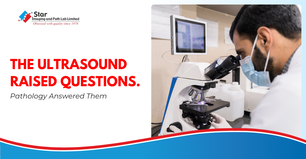
Microscopic evaluation revealed gastric mucosal tissue infiltrated by atypical round-to-ovoid cells arranged in nests and trabeculae. The cells exhibited finely stippled chromatin, moderate eosinophilic granular cytoplasm and mild cellular pleomorphism. These morphological features raised suspicion of a malignant neoplasm requiring further classification.
What Did the Histopathology Report Indicate?
The biopsy findings were suggestive of a poorly differentiated carcinoma, possibly a neuroendocrine tumour.
Poorly differentiated carcinomas are malignant tumours whose cells have lost many of the structural features associated with their normal tissue of origin. Identifying their precise type can be challenging because certain malignancies may share overlapping microscopic characteristics.
Histologically, GNETs exhibit diverse range of patterns, including solid, nested, trabecular, and glandular formations. IHC plays a critical role in establishing the diagnosis. GNETs typically shows positivity for markets such as S100, NSE(neuron-specific enolase), synaptophysin, and CD56. The expression of these neuroendocrine markers supports the neural crest origin of GNETs and helps distinguish them from other gastrointestinal tumors.
In this particular case, the microscopic appearance raised the possibility of neuroendocrine differentiation, but further testing was necessary before reaching a definitive classification.
Why Was Immunohistochemistry Recommended?
To investigate the suspected tumour further, the histopathology report recommended Immunohistochemistry (IHC).
IHC is a specialised laboratory technique that uses antibodies to identify specific proteins within tissue samples. The presence or absence of particular markers helps pathologists establish the cellular identity of a tumour and differentiate between malignancies with similar microscopic appearances.
For suspected neuroendocrine neoplasms, markers such as Synaptophysin, Chromogranin A and CD56 and NSE may be used, depending on the clinical and morphological findings. The Ki-67 proliferation index can also help assess how actively tumour cells are multiplying and contribute to tumour grading.
The choice of markers is determined by the pathologist based on the initial microscopic assessment. In this case, IHC was advised to establish whether the suspected malignancy demonstrated neuroendocrine differentiation and to support a more definitive diagnosis.
The Importance of Expert Pathological Evaluation
A complex biopsy requires more than identifying abnormal cells. It calls for careful assessment of their appearance, arrangement and distinguishing characteristics, followed by appropriate additional testing wherever necessary.
This case, evaluated by Dr. Abhilasha Maru, Consultant Pathologist at Star Imaging and Path Lab Limited, highlights the importance of a systematic approach to diagnosing unusual gastric lesions.
At Star Imaging and Path Lab Limited, histopathological assessment and appropriate ancillary investigations form an important part of evaluating tissue abnormalities and supporting clinical decision-making.
Ultimately, accurate tumour classification provides the treating clinician with essential diagnostic information for determining the subsequent course of evaluation and management.
An Expert Perspective
Dr. Abhilasha Maru
Consultant Pathologist, Star Imaging and Path Lab Limited
"In complex gastric biopsies, identifying malignant cells is only the first step. Establishing their precise nature and differentiation is equally important, particularly when the microscopic features overlap across different tumour types. Immunohistochemistry provides an additional layer of diagnostic evidence, allowing us to go beyond morphology and arrive at a more specific pathological classification. This precision is essential because the distinction between tumour categories can have significant implications for subsequent clinical management”.

FAQs
Question: What is a gastric biopsy and why is it performed?
Answer: A gastric biopsy involves collecting small tissue samples from the stomach lining, usually during an upper gastrointestinal endoscopy. These samples are examined under a microscope to investigate abnormalities such as nodules, ulcers, inflammation and suspected tumours. Histopathological examination helps establish the nature of the identified lesion.
Question: What does poorly differentiated carcinoma mean in a stomach biopsy report?
Answer: Poorly differentiated carcinoma refers to a malignant tumour whose cells show limited resemblance to normal tissue under microscopic examination. Additional investigations, including immunohistochemistry, may be required to establish the precise tumour type and differentiation.
Question: Can a gastric biopsy confirm a neuroendocrine tumour?
Answer: A gastric biopsy can raise suspicion of a neuroendocrine tumour through its microscopic appearance. However, confirmation generally requires correlation with immunohistochemical markers such as Synaptophysin and Chromogranin A, along with other appropriate investigations.
Question: What is the role of immunohistochemistry in gastric cancer diagnosis?
Answer: Immunohistochemistry uses specific antibodies to identify proteins within tissue samples. In suspected gastric malignancies, it can help distinguish between different tumour types, establish neuroendocrine differentiation and support more precise pathological classification.
Question: Which IHC markers are used to identify gastric neuroendocrine tumours?
Answer: Commonly used markers include Synaptophysin and Chromogranin A. INSM1 may also be considered in appropriate cases. The Ki-67 proliferation index helps assess tumour cell proliferation and contributes to grading. The final marker panel depends on the microscopic findings and suspected diagnosis.









Comments List