
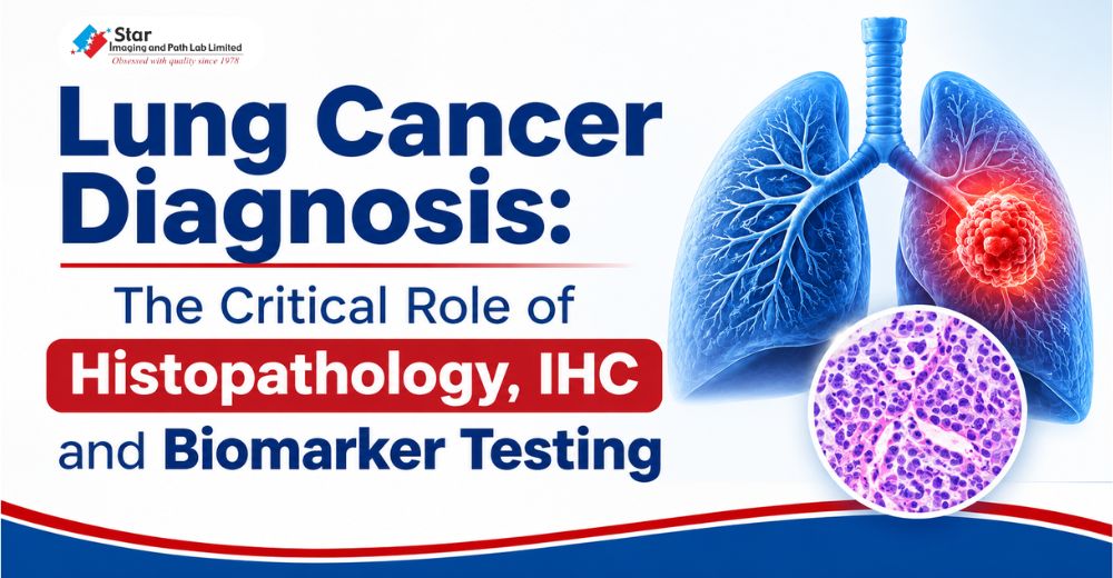
Lung Cancer Diagnosis: The Critical Role of Histopathology, IHC and Biomarker Testing
A lung cancer diagnosis raises an immediate concern, but it also opens up several important questions. What type of lung cancer is it? Are there specific changes within the tumour? And what information will help the treating doctor decide which treatment may be suitable?
These questions matter because lung cancer is not a single disease with one uniform pattern. Different types of lung cancer can behave differently, and even tumours belonging to the same broad category may have different molecular characteristics. This is why a diagnosis should go beyond simply confirming the presence of cancer.
A closer look at the tumour through Pathology and Laboratory investigations can provide information that may significantly influence treatment decisions.
Beyond the Diagnosis: What Does a Lung Cancer Pathology Report Really Tell Us?
When a doctor suspects lung cancer, imaging investigations can help identify an abnormal area and assess its location and extent. However, scans alone cannot establish the exact cancer type. For that, doctors generally need a tissue or cell sample, which is examined by a pathologist.
This examination is known as histopathology. The pathologist studies the cells under a microscope, looking at their appearance, arrangement and other features to determine whether cancer is present and, where possible, identify its specific type. Lung cancers are broadly divided into small cell lung cancer (SCLC) and non-small cell lung cancer (NSCLC), with NSCLC further classified into subtypes such as adenocarcinoma and squamous cell carcinoma.
Why does this distinction matter? Because the treatment approach is influenced by the type of lung cancer, its stage and other clinical findings. A more specific diagnosis gives the treating team a better starting point for planning the next steps.
When the Microscope Needs a Little More Help: The Role of IHC Testing
Sometimes, the appearance of cancer cells does not provide enough information to classify a tumour confidently. In such situations, pathologists may recommend Immunohistochemistry (IHC) testing.
IHC uses specially selected antibodies to detect particular proteins in tissue samples. These proteins act like markers that can help distinguish one type of tumour from another. For example, certain IHC markers can help differentiate lung adenocarcinoma from squamous cell carcinoma or help investigate whether a tumour has originated in the lung or spread from another organ.
In simple terms, histopathology helps examine what the cells look like, while IHC provides additional clues about what those cells are. Together, they can make lung cancer diagnosis more precise.
Biomarker Testing: Looking for Clues That Can Influence Treatment
Identifying the type of lung cancer is important, but there is another layer of information that may be relevant, particularly in non-small cell lung cancer.
Cancer cells can carry specific genetic changes or express certain proteins that may influence how the disease responds to treatment. These are known as biomarkers.
Lung cancer biomarker testing looks for such changes. Depending on the cancer type and clinical situation, testing may include EGFR, ALK, ROS1, BRAF, KRAS, MET, RET and other relevant markers. PD-L1 testing is another important assessment that can help doctors evaluate whether certain immunotherapy options may be appropriate.
The findings can help identify patients who may benefit from targeted therapy or immunotherapy. For instance, identifying an actionable EGFR alteration may help the oncologist consider an EGFR-targeted treatment.
This is one of the reasons molecular testing has become an important part of personalised lung cancer care. Instead of looking only at where the cancer has developed, doctors can also consider the biological characteristics of the tumour.
Why Histopathology, IHC and Biomarker Testing Need to Work Together
These investigations answer different questions, but their findings contribute to the same clinical picture. Histopathology establishes the diagnosis and helps classify the cancer. IHC adds clarity when further distinction is needed, while biomarker testing looks for molecular or protein changes that may have treatment implications.
Not every patient requires every test. The investigations recommended depend on the type and stage of cancer, the available tissue sample and the treating oncologist's clinical assessment. Making the best use of the available sample is also important, as sufficient tissue may be needed for several stages of testing.
At Star Imaging and Path Lab Limited, we recognise that pathology is not simply about identifying abnormal cells. It is about generating reliable, meaningful diagnostic information that helps clinicians understand the disease more clearly.
As we observe Lung Cancer Awareness Month, it is worth remembering that an accurate diagnosis involves more than detecting cancer. Understanding its type, characteristics and relevant biomarkers can provide valuable information for the decisions that follow.
Because every cancer diagnosis deserves a closer look, not just a confirmation.
FAQs
Question: Can a CT scan confirm whether a person has lung cancer?
Answer: A CT scan can reveal suspicious growths, nodules or other abnormalities in the lungs, helping doctors identify areas that need further investigation. However, a scan alone cannot confirm the exact type of cancer. A tissue or cell sample is generally examined through pathological testing to establish the diagnosis and classify the tumour.
Question: What is the difference between a histopathology test and an IHC test in lung cancer?
Answer: Histopathology involves examining tissue under a microscope to identify cancer cells and understand their appearance and arrangement. IHC, or Immunohistochemistry, uses specific antibodies to detect proteins within the tissue. These protein markers provide additional information that can help pathologists distinguish between different tumour types when the initial examination is not conclusive.
Question: Why is biomarker testing important after a lung cancer diagnosis?
Answer: Biomarker testing looks for specific genetic changes or protein markers in cancer cells. Certain findings can help doctors identify whether a patient may be suitable for targeted therapy or immunotherapy. It provides information beyond the cancer's location and type, supporting a more individualised approach to treatment planning.
Question: Which biomarkers are tested in non-small cell lung cancer?
Answer: Depending on the cancer subtype, stage and clinical requirements, testing may include EGFR, ALK, ROS1, BRAF, KRAS, MET, RET and other relevant biomarkers. PD-L1 is also commonly assessed through IHC testing because its expression can help guide decisions about certain immunotherapy options. The exact testing panel is determined by the treating team.
Question: Does every lung cancer patient need IHC and molecular biomarker testing?
Answer: Not necessarily. The need for additional investigations depends on the type of lung cancer, its stage, the initial pathology findings and the treatment being considered. IHC may be recommended to establish a more specific diagnosis, while molecular biomarker testing is particularly important in treatment planning for eligible patients with non-small cell lung cancer. The treating oncologist and pathologist determine which tests are appropriate.







Comments List